We are the ONLY providers in the tri-state area using microscopes to perform root canals. We believe the microscope is essential to the long term success of a root canal.
Our doctors use very specialized equipment:
Dental Operating Microscope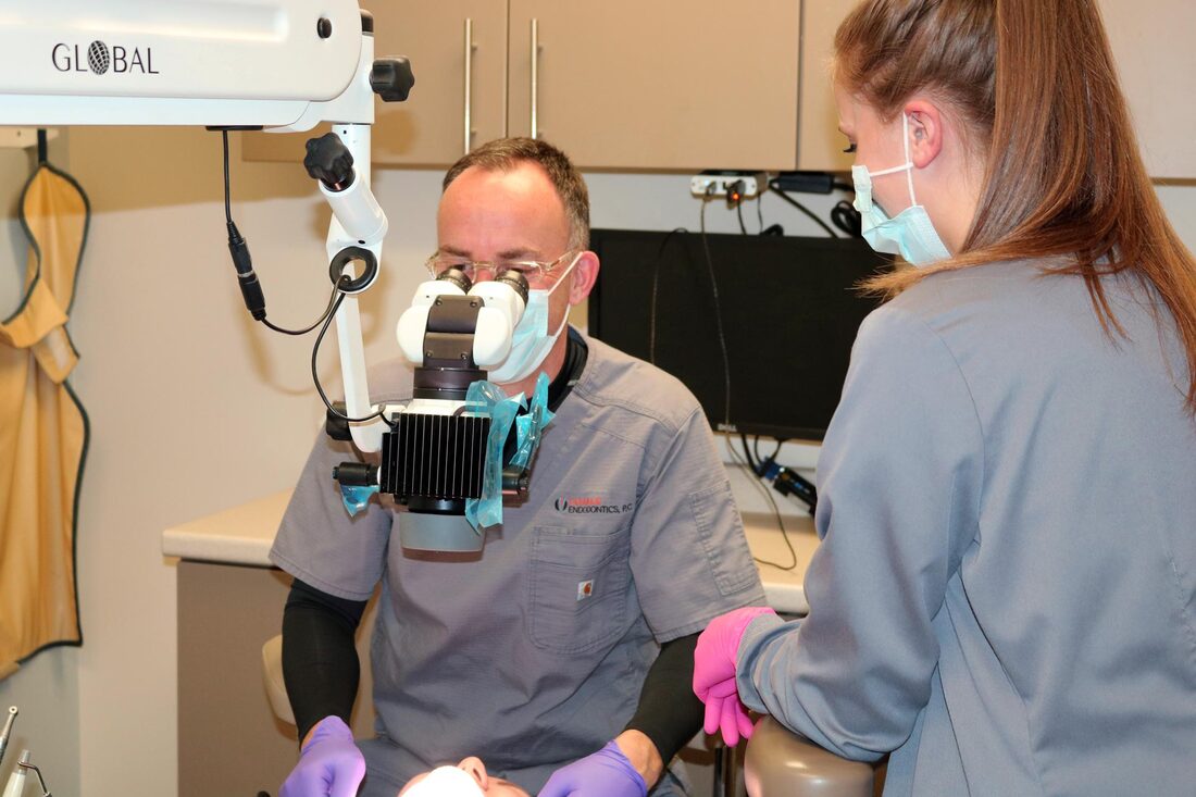
General dentists typically wear glasses with magnifying lenses attached to them. These are called loupes, and they can magnify the tooth from 2.5 to 4.5 times the original size. Because nerves in the tooth are so incredibly small, endodontists rely on a much higher degree of magnification to be able to find them. We use a dental operating microscope to magnify canals/nerves up to 25 times the original size! This allows for a level of accuracy that is only possible through use of this technology. Our microscope hangs from the ceiling and we look through it during treatment. It has internal lenses and an LED light source that allows us to visualize canals/nerves that are often missed during non-microscope root canal treatments. It can also allow us to see cracks, decay, and small defects that can lead to root canal failure if not recognized and managed. Most importantly, the use of the microscope enables us to remove the least amount of tooth structure possible. The more tooth we can preserve, the longer you will keep your tooth. We are the ONLY providers in the tri-state area using microscopes to perform root canals. |
Cone Beam Computed Tomography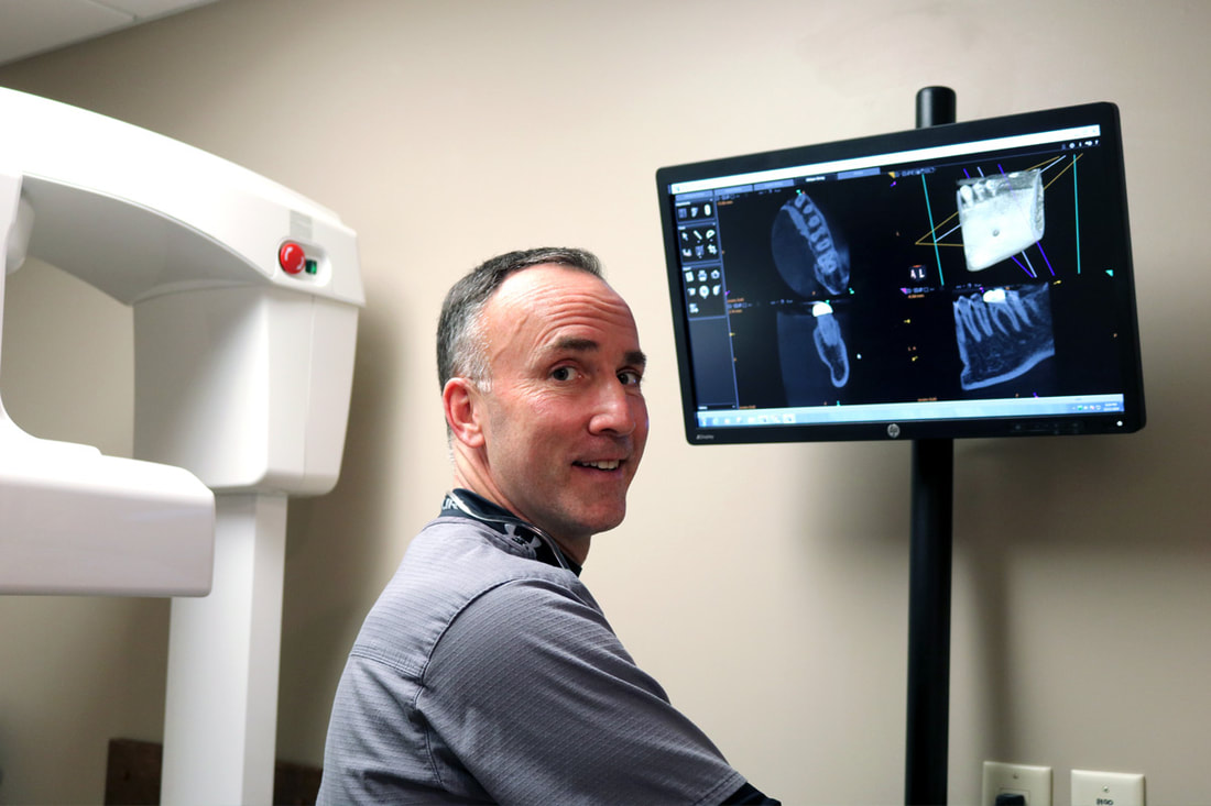
Our office also utilizes the most advanced imaging possible. Cone beam computed tomography (CBCT) imaging is heavily utilized by endodontists around the country. CBCT imaging is similar to a CT scan taken in a medical office, but with a much smaller field of view (four cubic centimeters). The CBCT scan is not used for all cases. In many treatments the standard two-dimensional (2-D) dental x-ray is sufficient. However, when a three-dimensional (3-D) view of the tooth and surrounding tissue is needed, we have the ability to acquire it. The 3-D scan is useful for the detection of abscesses not easily viewed in 2-D, such as maxillary (upper) molars whose roots are often surrounded by maxillary sinus. The 3-D scan is often used to diagnose fractured roots prior to treatment which enables us to limit unneeded root canal procedures. Most importantly, the scan taken preoperatively or mid-treatment helps identify the complex anatomy of many teeth. This anatomy includes the fourth canal in three-rooted maxillary molars, and the lingual canal of mandibular (lower) incisors and premolars. Why is this important? Well, not all teeth are alike, some have extra canals, others do not. If one or more of the canals are not found, then the treatment will more than likely fail. These advanced technologies enable us to quickly and effectively complete your root canal treatment. |
|
(563) 583-1050
[email protected] 988 W. 3rd Street, Suite 107 Dubuque IA 52001 Office Hours: Monday-Wednesday 7:30am-4:30pm Thursday 7:30am-4:00pm |


©
2020-2022 Tri State Root Canal Specialists | Privacy Policy | Terms Of Service
Website by LislDesign.com
Website by LislDesign.com